
CilOCT: A software for semi-automatic segmentation and analysis of the ciliary muscle in OCT images
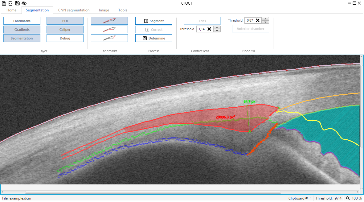
CilOCT is an open-source software for semi-automatic segmentation of the ciliary muscle (CM) within optical coherence tomography (OCT) images. It provides direct import of OCT images in DICOM format, a standardized procedure for segmentation, image distortion correction, the export of CM landmarks, like ciliary muscle apex and scleral spur, as well as a continuous thickness profile of the ciliary muscle as a novel way of analysis. Additionally, it supports batch processing for the automated analysis of large numbers of images.
Software
Publications
- Sandra Wagner, Eberhart Zrenner, and Torsten Strasser. 2018. “Ciliary Muscle Thickness Profiles Derived from Optical Coherence Tomography Images.” Biomedical Optics Express 9 (10): 5100. doi:10.1364/BOE.9.005100.
- Sandra Wagner, Eberhart Zrenner, and Torsten Strasser. 2019. “Emmetropes and Myopes Differ Little in Their Accommodation Dynamics but Strongly in Their Ciliary Muscle Morphology.” Vision Research 163 (October). Elsevier: 42–51. doi:10.1016/j.visres.2019.08.002.
- Sandra Wagner, Frank Schaeffel, Eberhart Zrenner, and Torsten Straßer. 2019. “Prolonged Nearwork Affects the Ciliary Muscle Morphology.” Experimental Eye Research 186 (September): 107741. doi:10.1016/j.exer.2019.107741.
- Torsten Straßer, Sandra Wagner, and Eberhart Zrenner. 2020. “Review of the Application of the Open-Source Software CilOCT for Semi-Automatic Segmentation and Analysis of the Ciliary Muscle in OCT Images.” Edited by Asaf Achiron. PLOS ONE 15 (6): e0234330. doi:10.1371/journal.pone.0234330.